Your bathroom scale tells you one story. The mirror tells you another. But neither reveals what might be the most important health metric you’re ignoring: the fat you can’t see, wrapped around your liver, kidneys, and intestines, quietly damaging your arteries while you maintain what looks like a healthy weight.
A groundbreaking 2025 study from McMaster University, analyzing MRI scans of over 33,000 adults, has confirmed what researchers have long suspected. Deep abdominal fat and liver fat can silently damage your cardiovascular system even when your BMI falls squarely in the “normal” range. The implications are significant: millions of people who consider themselves healthy based on weight alone may be harboring metabolic dysfunction that traditional metrics completely miss.
This isn’t about body shaming or aesthetic preferences. It’s about understanding that body composition, specifically where your body stores fat, matters far more than the number on your scale. The good news is that visceral fat responds remarkably well to targeted lifestyle interventions, often more readily than the stubborn subcutaneous fat most people obsess over.
What Makes Visceral Fat Different
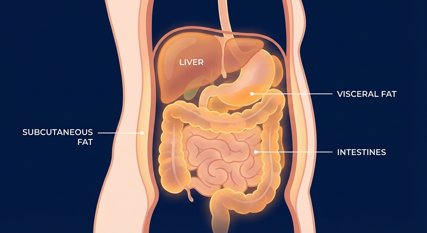
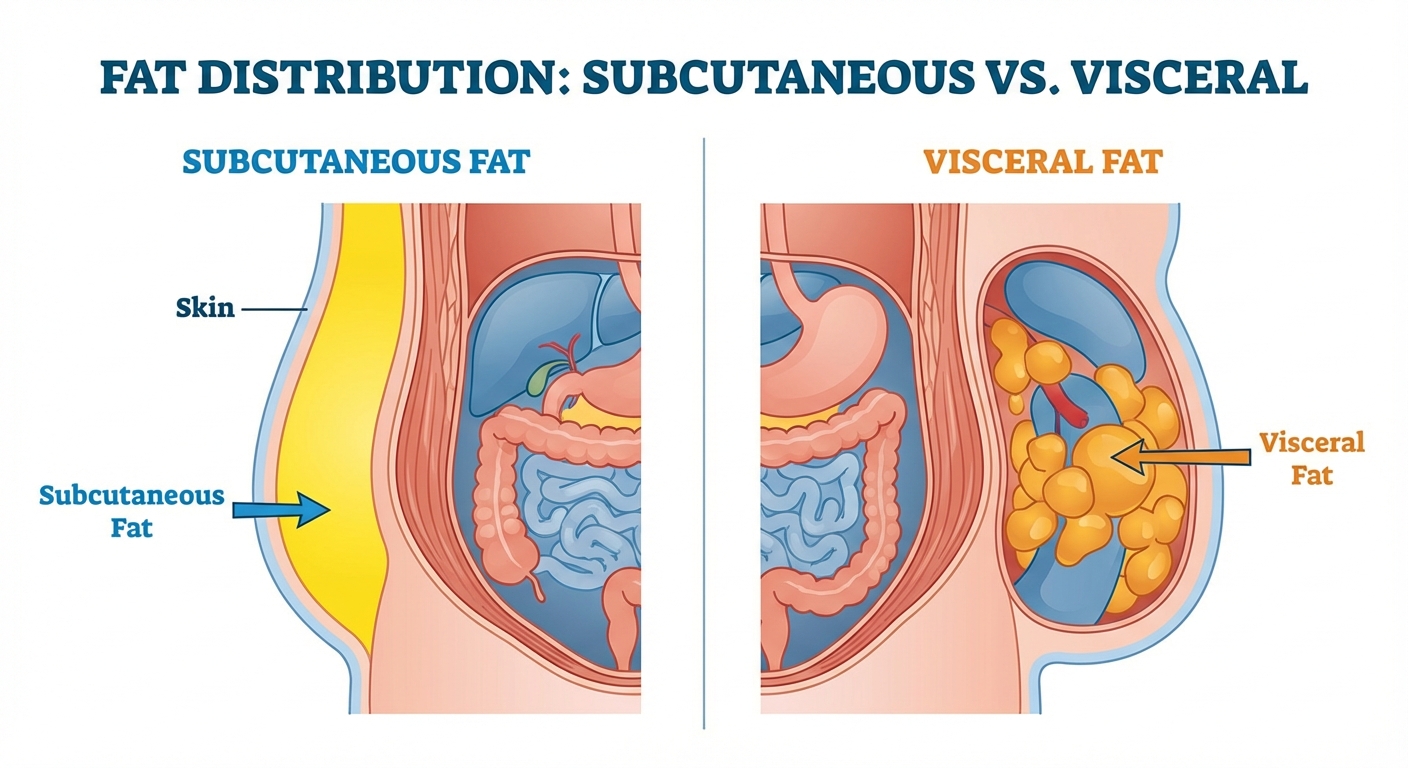
Not all body fat functions the same way. Subcutaneous fat, the kind you can pinch between your fingers, sits directly beneath your skin and serves primarily as energy storage and insulation. While excess subcutaneous fat carries some health risks, it’s relatively metabolically inactive compared to its more dangerous counterpart.
Visceral fat, also called intra-abdominal fat, occupies the spaces between and around your internal organs. Your liver, pancreas, intestines, and kidneys can all become surrounded by this metabolically active tissue. Unlike subcutaneous fat, visceral fat doesn’t just passively store calories. It actively secretes hormones, inflammatory compounds, and signaling molecules that directly influence your metabolism, immune function, and cardiovascular health.
The liver deserves special attention here. When fat accumulates within liver cells, a condition called hepatic steatosis or fatty liver disease, it disrupts the organ’s ability to regulate blood sugar, process cholesterol, and filter toxins. The McMaster research found that liver fat showed particularly strong associations with arterial stiffness, a precursor to cardiovascular disease, even in participants with otherwise healthy-looking body compositions.
What makes visceral fat so metabolically dangerous? The fat cells in this region have direct venous drainage to the liver through the portal vein, meaning inflammatory compounds and fatty acids released by visceral fat tissue get first-pass exposure to your liver before circulating through the rest of your body. This anatomical quirk explains why visceral fat accumulation so strongly predicts metabolic dysfunction, liver disease, and cardiovascular problems.
The “Thin Outside, Fat Inside” Phenomenon
The McMaster study brought renewed attention to a concept that metabolic researchers have been discussing for years: the TOFI phenotype, or “thin outside, fat inside.” These individuals maintain normal body weight and may even appear lean, yet harbor significant visceral and liver fat deposits that create metabolic profiles similar to people with obvious obesity.
TOFI individuals often have normal BMIs but elevated waist circumferences, suggesting their body preferentially stores fat around organs rather than under the skin. They may pass standard medical screenings with flying colors while their arteries silently stiffen and their metabolic health deteriorates. The McMaster research found that even modest increases in visceral and liver fat correlated with measurable arterial changes in participants who would never be flagged as at-risk based on weight alone.
The phenomenon appears more common than previously thought. Some estimates suggest up to 30% of normal-weight adults carry metabolically significant visceral fat deposits. Genetics play a role, as does age, hormonal status, and lifestyle factors. Men tend to accumulate visceral fat more readily than premenopausal women, though this pattern shifts after menopause when women’s visceral fat accumulation accelerates.
Perhaps most concerning, TOFI individuals often feel a false sense of security about their health. They don’t receive the warnings that visibly overweight people get from doctors, family members, or their own reflection. They may even engage in unhealthy behaviors under the assumption that their weight protects them. The McMaster findings suggest this assumption is dangerously misguided.
How Visceral Fat Damages Your Cardiovascular System
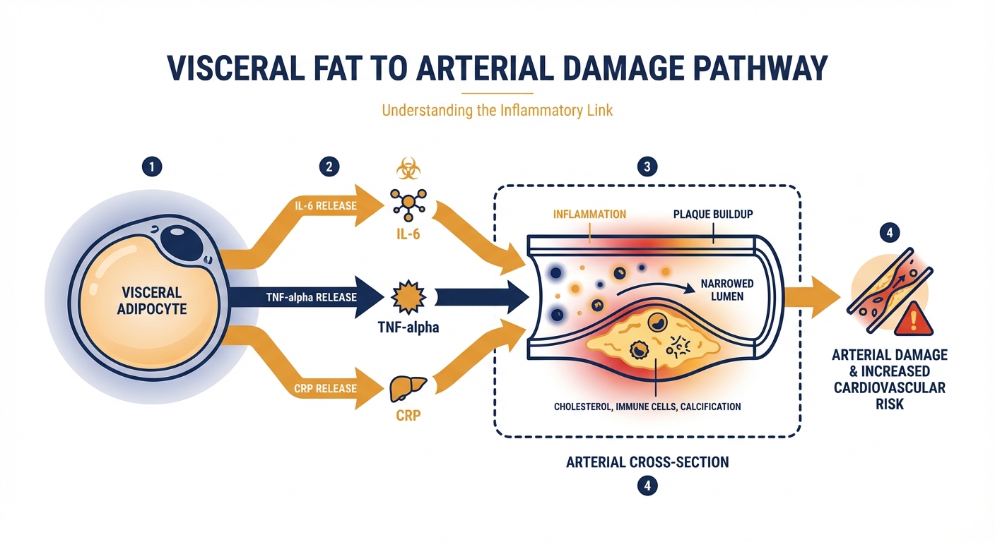
The pathway from visceral fat to cardiovascular disease involves multiple interconnected mechanisms. Understanding these pathways helps explain why this particular fat depot carries such outsized health risks and informs strategies for intervention.
Chronic low-grade inflammation sits at the center of visceral fat’s harmful effects. Visceral adipose tissue secretes pro-inflammatory cytokines, including interleukin-6 (IL-6), tumor necrosis factor-alpha (TNF-α), and C-reactive protein (CRP). These inflammatory mediators damage blood vessel walls, promote plaque formation, and interfere with insulin signaling throughout the body. The McMaster researchers noted that inflammatory markers tracked closely with both visceral fat volume and arterial stiffness measurements.
Insulin resistance represents another critical mechanism. When visceral fat releases excess free fatty acids into the bloodstream, muscle and liver cells become less responsive to insulin’s signals. The pancreas compensates by producing more insulin, creating a state of hyperinsulinemia that further promotes fat storage, inflammation, and arterial damage. This vicious cycle can persist for years before blood sugar levels rise enough to trigger a diabetes diagnosis, meaning the damage accumulates silently.
Visceral fat also disrupts the balance of adipokines, hormones produced by fat tissue that regulate appetite, metabolism, and vascular health. Healthy fat tissue produces adequate adiponectin, a protective hormone that enhances insulin sensitivity and reduces inflammation. Visceral fat accumulation suppresses adiponectin production while increasing leptin resistance, creating hormonal imbalances that perpetuate metabolic dysfunction and cardiovascular risk.
Assessing Your Visceral Fat Risk
While MRI scans provide the most accurate assessment of visceral fat, several practical measurements can help you gauge your risk without expensive imaging.
Waist circumference remains one of the most reliable and accessible indicators. For men, a waist measurement above 40 inches (102 cm) indicates elevated visceral fat risk. For women, the threshold sits at 35 inches (88 cm). Measure at the level of your navel, not at your belt line or the narrowest part of your waist. Take the measurement first thing in the morning before eating, with your abdominal muscles relaxed.
Waist-to-hip ratio provides additional insight by accounting for body frame differences. Divide your waist circumference by your hip circumference, measured at the widest point of your buttocks. Ratios above 0.90 for men or 0.85 for women suggest visceral fat accumulation that warrants attention. This metric often catches TOFI individuals who might have acceptable waist measurements but disproportionate abdominal fat relative to their overall body composition.
Waist-to-height ratio has emerged as another useful screening tool. Simply divide your waist circumference by your height. A ratio above 0.5 indicates elevated risk regardless of your weight or BMI. This measurement works across different body types and ethnicities, making it particularly valuable for populations where standard BMI cutoffs may not accurately reflect metabolic risk.
Blood markers can supplement physical measurements. Elevated fasting triglycerides, especially when combined with low HDL cholesterol, strongly correlate with visceral fat accumulation. Elevated fasting insulin or a high HOMA-IR score, which estimates insulin resistance, also suggest visceral fat may be affecting your metabolic health. These markers often become abnormal before weight gain becomes visible or BMI crosses into concerning territory.
Evidence-Based Strategies for Reducing Visceral Fat
The encouraging news from metabolic research: visceral fat often responds more readily to intervention than subcutaneous fat. While you can’t spot-reduce fat from any area through targeted exercise, certain lifestyle modifications preferentially mobilize visceral fat stores.
Aerobic exercise shows particularly strong effects on visceral fat reduction. A meta-analysis of 117 studies found that moderate-intensity continuous training, equivalent to brisk walking or easy cycling for 30-60 minutes most days, reduced visceral fat even when total body weight remained stable. The mechanism involves increased fatty acid oxidation and improved insulin sensitivity in visceral adipose tissue. Zone 2 cardio, performed at an intensity where you can still hold a conversation, appears especially effective for metabolic health improvements.
High-intensity interval training (HIIT) may produce even faster visceral fat reductions in less time. Studies comparing HIIT to moderate-intensity exercise have found greater visceral fat loss with HIIT despite lower total exercise time and similar caloric expenditure. The metabolic stress of high-intensity efforts appears to preferentially target visceral fat stores and improve insulin sensitivity more powerfully than steady-state cardio alone.

Resistance training contributes to visceral fat reduction through multiple pathways. Building muscle mass increases your basal metabolic rate, meaning you burn more calories at rest. Resistance exercise also improves insulin sensitivity and glucose uptake in muscle tissue, reducing the hormonal signals that promote visceral fat storage. The combination of resistance training and aerobic exercise appears more effective than either modality alone for reducing visceral fat and improving metabolic markers.
Dietary modifications can accelerate visceral fat loss. Reducing refined carbohydrates and added sugars helps lower insulin levels, creating hormonal conditions that favor visceral fat mobilization. Adequate protein intake, around 1.6-2.2 grams per kilogram of body weight, supports muscle preservation during fat loss and enhances satiety. Mediterranean-style eating patterns, emphasizing vegetables, olive oil, nuts, fish, and modest wine consumption, have shown consistent benefits for reducing both visceral fat and cardiovascular risk markers.
Time-restricted eating, limiting food consumption to an 8-10 hour daily window, may preferentially target visceral fat. Studies have shown greater reductions in waist circumference and liver fat with time-restricted eating compared to calorie-matched diets without time constraints. The mechanism likely involves improved circadian alignment and extended periods of lower insulin levels that allow visceral fat mobilization.
Sleep, Stress, and Visceral Fat Accumulation
Lifestyle factors beyond diet and exercise significantly influence where your body stores fat. Chronic sleep deprivation and elevated stress hormones both promote visceral fat accumulation, even when caloric intake remains constant.
Sleep restriction alters hormonal patterns in ways that favor visceral fat storage. Just one week of sleeping 5 hours per night, instead of 8, increases visceral fat accumulation by approximately 9% according to Mayo Clinic research. The mechanism involves disrupted cortisol rhythms, reduced growth hormone secretion, and impaired insulin sensitivity. Prioritizing 7-9 hours of quality sleep supports healthy body composition and may be as important as exercise for managing visceral fat.
Chronic stress elevates cortisol levels, and cortisol strongly promotes visceral fat deposition. The stress hormone specifically activates enzymes in visceral fat tissue that convert inactive cortisone to active cortisol, creating a local amplification effect. Stress management techniques, including evidence-based breathing practices and vagal nerve activation, can help interrupt this cycle and reduce the hormonal drive toward visceral fat storage.
Alcohol consumption deserves mention given its direct effects on liver fat and visceral fat accumulation. Even moderate drinking, defined as one drink daily for women or two for men, increases liver fat deposition. Higher consumption levels strongly promote both liver fat and visceral adiposity, with the classic “beer belly” representing visible evidence of alcohol’s effects on abdominal fat distribution. Reducing or eliminating alcohol often produces noticeable improvements in waist circumference and metabolic markers within weeks.
The Bottom Line
The McMaster research confirms what body composition experts have long understood: your weight tells only a small part of your metabolic story. Visceral fat, hidden deep in your abdomen, can quietly damage your cardiovascular system while you maintain what appears to be a healthy weight. Traditional metrics like BMI miss this hidden risk entirely, leaving millions of TOFI individuals unaware of their true metabolic status.
The practical takeaway isn’t to obsess over another number, but to recognize that body composition matters more than body weight. Simple measurements like waist circumference and waist-to-hip ratio provide meaningful insights without expensive imaging. Blood markers including triglycerides, HDL cholesterol, and fasting insulin can further clarify your metabolic picture.
Next Steps:
- Measure your waist circumference at navel level first thing tomorrow morning
- Calculate your waist-to-height ratio, and flag readings above 0.5 for further investigation
- Request fasting triglycerides and insulin levels at your next blood draw
- Prioritize Zone 2 cardio and resistance training, which preferentially reduce visceral fat
- Address sleep quality and stress management as seriously as diet and exercise
Sources: McMaster University MRI study (2025), Mayo Clinic sleep and visceral fat research, Journal of Clinical Endocrinology & Metabolism visceral fat mechanisms review, American Heart Association body composition guidelines.