The MRI scan reveals everything. Not just the current state of your brain, but how old it looks compared to your actual age. For some people in the study, their brains appeared years younger than their birth certificates suggested. For others, the opposite was true. The question researchers set out to answer wasn’t whether this variation existed, but what explained it. The answer, it turns out, had less to do with brain-specific interventions and more to do with what was happening below the neck.
A major study presented at the Radiological Society of North America’s annual meeting this November analyzed whole-body MRI scans from 1,164 healthy adults and found a striking pattern. Those with higher muscle mass and lower visceral fat showed significantly younger brain ages than their chronological years would predict. The fat just under the skin, the kind you can pinch, showed no relationship to brain aging. But the deep abdominal fat surrounding internal organs, combined with muscle volume, told a different story entirely.
Dr. Cyrus Raji, associate professor of radiology and neurology at Washington University School of Medicine in St. Louis, led the research team. His conclusion was direct: “More muscle and a lower visceral fat to muscle ratio were linked to a younger brain.” The implications ripple outward from neurology into practical fitness advice, weight loss medication design, and how we conceptualize the relationship between physical training and cognitive longevity.
The Study: What 1,164 Brain Scans Revealed
The research team examined healthy adults across four sites, with an average age of 55.17 years. Women comprised 52% of the participants. Each underwent comprehensive MRI imaging using T1-weighted sequences, a technique that produces images where fat appears bright and fluid appears dark, allowing precise quantification of different tissue types.
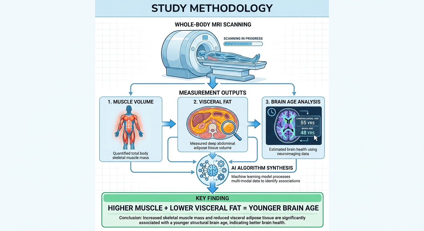
An artificial intelligence algorithm trained on 5,500 independent brain MRI scans calculated biological brain age for each participant. This AI model identifies structural characteristics that correlate with aging, essentially comparing each brain’s appearance to what would be expected for someone of that chronological age. The gap between predicted brain age and actual age became the key variable.
Separately, the AI quantified total normalized muscle volume, visceral fat (the metabolically active fat surrounding abdominal organs), and subcutaneous fat (the surface fat beneath the skin). The research question was whether these body composition variables predicted brain aging independent of other factors.
The results were unambiguous. Participants with higher visceral-to-muscle ratios displayed older predicted brain ages. Those with greater muscle mass showed younger-appearing brains. Subcutaneous fat, despite its cosmetic prominence, showed no meaningful correlation with brain aging. The distinction matters because it identifies which body composition changes might actually influence brain health versus which are merely aesthetic concerns.
Why Muscle Protects the Brain
The mechanisms connecting muscle tissue to brain health operate through multiple pathways, and understanding them transforms resistance training from a vanity pursuit into a neuroprotective strategy.
Muscle tissue is metabolically active in ways that influence systemic health. Skeletal muscle serves as the primary site for glucose disposal after meals, meaning more muscle mass creates greater metabolic sink capacity for blood sugar. Poor glucose regulation is strongly associated with accelerated brain aging, cognitive decline, and increased dementia risk. By improving metabolic health, muscle mass may protect the brain from the damage that chronically elevated blood sugar causes.
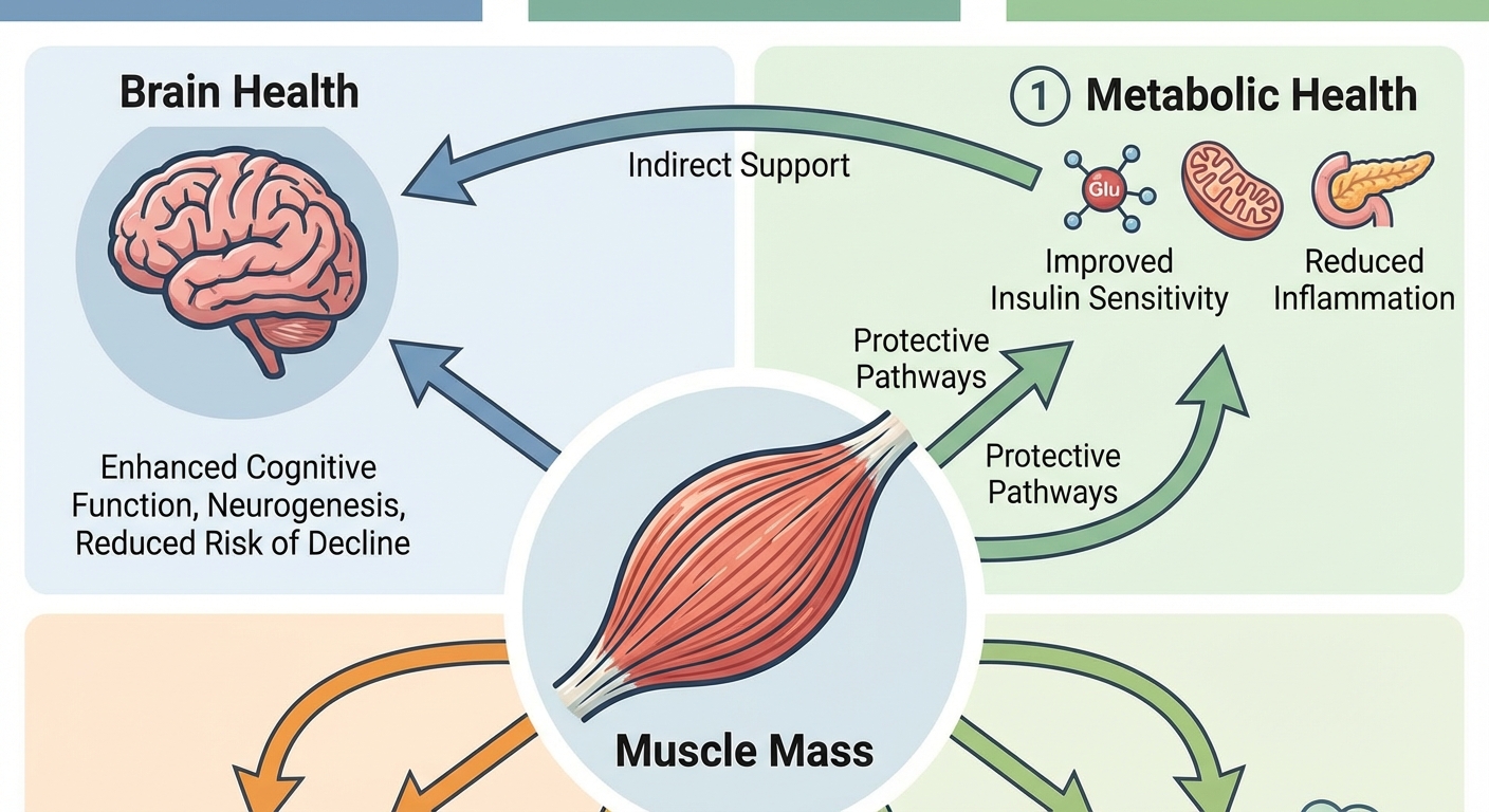
Muscle also releases myokines, signaling molecules that function similarly to hormones. Among these, brain-derived neurotrophic factor (BDNF) has garnered particular attention. BDNF promotes neuroplasticity, supports the survival of existing neurons, and encourages the growth of new neurons and synapses. Exercise, particularly resistance training that challenges muscles, increases BDNF production. The more muscle you have engaged in regular activity, the more of this neuroprotective signal you generate.
Inflammation provides another connection point. Visceral fat is not inert tissue. It actively secretes inflammatory cytokines that circulate throughout the body, crossing the blood-brain barrier and contributing to neuroinflammation. Chronic low-grade inflammation accelerates brain aging and increases risk for neurodegenerative diseases. Muscle tissue, in contrast, tends to be anti-inflammatory when regularly exercised. The ratio of muscle to visceral fat thus influences systemic inflammatory load, with downstream effects on brain health.
Dr. Raji emphasized this point: “Having more muscle and less visceral fat is better for your metabolic health, which promotes a healthier brain. The combination also lowers inflammation in the brain, which can reduce the impact of aging.”
The Visceral Fat Problem
Not all body fat is created equal, and this study reinforces a distinction that metabolic researchers have emphasized for decades. Visceral fat, the type that accumulates deep in the abdominal cavity around organs like the liver, pancreas, and intestines, behaves fundamentally differently from the subcutaneous fat that sits just beneath the skin.
Visceral fat functions almost like an endocrine organ, releasing inflammatory compounds, disrupting insulin signaling, and contributing to metabolic syndrome. It’s strongly associated with cardiovascular disease, type 2 diabetes, and, as this research confirms, accelerated brain aging. The problem is that visceral fat isn’t always visible. Thin individuals can carry significant visceral fat deposits, a phenomenon sometimes called “skinny fat” or more technically, metabolically obese normal weight.
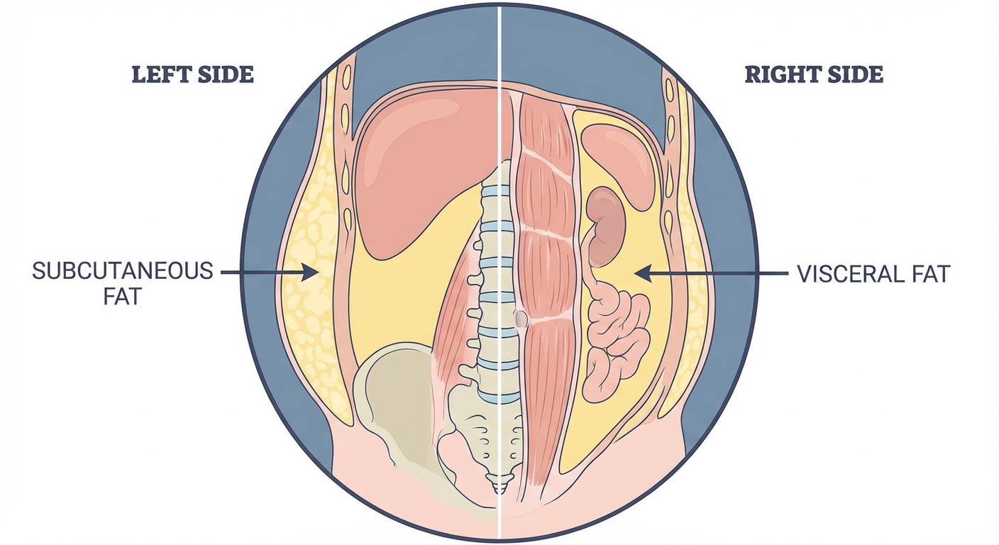
The MRI imaging used in this study allowed researchers to precisely quantify visceral fat separate from subcutaneous fat, something not possible with standard body composition assessments like BMI or even basic body fat percentage measurements. This precision revealed that only visceral fat showed the concerning association with brain aging, while the fat that concerns most people when they look in the mirror, the subcutaneous variety, showed no relationship.
This finding has practical implications. Fat loss strategies that reduce subcutaneous fat without addressing visceral fat may improve appearance without improving brain health. Conversely, interventions that specifically target visceral fat, even if they don’t dramatically change appearance, may offer significant neuroprotective benefits.
Implications for GLP-1 Weight Loss Medications
The research arrives at a moment when GLP-1 receptor agonist medications like Ozempic and Wegovy have revolutionized weight loss treatment. These drugs produce significant fat loss, but they also cause muscle loss, a phenomenon that has concerned some clinicians and researchers.
Dr. Raji addressed this directly: “While widely prescribed GLP-1 weight loss drugs are powerful at inducing fat loss, they may also be related to a higher burden of muscle loss.” Given that muscle mass appears protective for brain health while visceral fat appears harmful, the composition of weight loss matters, not just the total pounds lost.
The ideal scenario based on this research would be losing visceral fat while preserving or building muscle. “Losing fat, especially visceral fat, while preserving muscle volume would have the best benefit on brain aging and brain health based on insights from our work,” Raji explained.
This has immediate practical implications for anyone using weight loss medications. Resistance training during GLP-1 treatment becomes not just advisable but potentially essential for preserving the brain health benefits that should accompany improved metabolic health. Without deliberate muscle preservation efforts, the net effect of dramatic weight loss on brain aging could be less favorable than expected. For more on optimizing protein intake during GLP-1 treatment, see our guide on protein timing for GLP-1 users.
Building the Brain-Protective Body
Translating these findings into action requires understanding what actually builds muscle and reduces visceral fat. The good news is that both goals respond to similar interventions, though with different emphasis.
Resistance training is the primary driver of muscle mass. This doesn’t require becoming a bodybuilder. Moderate resistance training two to three times weekly, progressively challenging muscles with weights, machines, or bodyweight exercises, stimulates the adaptations that build and maintain muscle tissue. For adults over 50, our guide on strength training after 50 provides specific protocols optimized for the aging body. The key is progressive overload: gradually increasing demands over time to continually stimulate adaptation.
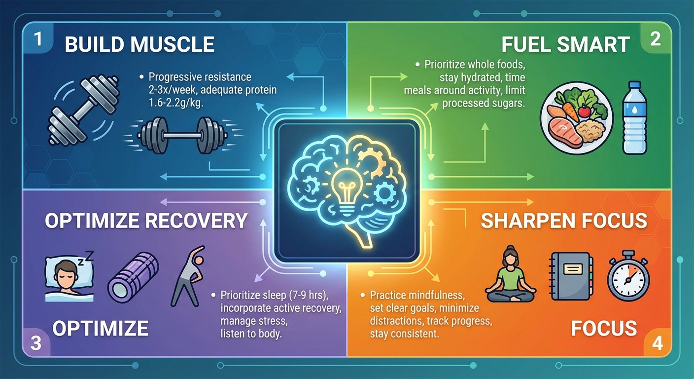
Protein intake supports muscle maintenance and growth. Current research suggests 1.6 to 2.2 grams of protein per kilogram of body weight daily for individuals engaged in resistance training, with higher intakes particularly important for older adults and those in caloric deficit. Distribution matters too: spreading protein intake across meals rather than concentrating it in one sitting optimizes muscle protein synthesis throughout the day.
Visceral fat responds particularly well to consistent moderate activity, dietary changes that reduce refined carbohydrates and processed foods, adequate sleep (insufficient sleep increases cortisol, which promotes visceral fat deposition), and stress management (chronic stress similarly elevates cortisol). Unlike subcutaneous fat, which can be stubborn, visceral fat often responds relatively quickly to lifestyle intervention.
The combination of resistance training and these lifestyle factors creates a synergistic effect. Resistance training itself reduces visceral fat independent of caloric deficit, while the muscle it builds improves metabolic health in ways that further discourage visceral fat accumulation. The goal isn’t just weight management but body composition optimization: more muscle, less visceral fat, regardless of what the scale says.
Measuring What Matters
Standard health metrics don’t capture the body composition factors this research identifies as important. BMI, the most commonly used measure, cannot distinguish between muscle and fat, meaning a muscular person and an overfat person of the same height and weight will have identical BMIs despite dramatically different health profiles.
Body fat percentage moves closer to what matters but doesn’t distinguish between visceral and subcutaneous fat. DEXA scans, increasingly available in fitness-focused medical practices, provide detailed body composition data including estimates of visceral fat. Whole-body MRI, as used in this study, offers the most precise assessment but remains expensive and not widely available.
A practical proxy for visceral fat is waist circumference. Because visceral fat accumulates in the abdominal cavity, waist measurement correlates reasonably well with visceral fat volume. Waist circumference above 40 inches in men or 35 inches in women indicates elevated visceral fat risk. The waist-to-hip ratio provides additional information: higher ratios suggest proportionally more central fat, which tends to be visceral.
For muscle mass, strength itself serves as a reasonable proxy. If you’re getting stronger over time, you’re likely building or maintaining muscle. Grip strength, easily measured with an inexpensive dynamometer, correlates with overall muscle mass and predicts longevity independent of other factors. Performance on functional tests like the ability to rise from a seated position without using hands also reflects muscle health.
The Bottom Line
A major study using whole-body MRI on 1,164 adults found that higher muscle mass and lower visceral fat both independently correlate with younger-appearing brains. The research, led by Dr. Cyrus Raji at Washington University School of Medicine, suggests that body composition may influence brain aging through metabolic, inflammatory, and neurotrophic mechanisms. Importantly, subcutaneous fat showed no relationship to brain age, highlighting that the type of fat, not just the amount, matters for brain health.
These findings have immediate practical implications: resistance training and lifestyle factors that build muscle while reducing visceral fat may offer neuroprotective benefits beyond their metabolic effects. For those using GLP-1 weight loss medications, preserving muscle through resistance training and adequate protein becomes particularly important to ensure that weight loss benefits brain health rather than potentially compromising it.
Next Steps:
- Incorporate resistance training 2-3 times weekly, focusing on progressive overload
- Ensure adequate protein intake (1.6-2.2g per kg body weight daily, distributed across meals)
- Measure waist circumference as a proxy for visceral fat; target under 40 inches for men, under 35 inches for women
- Prioritize sleep and stress management, both of which influence visceral fat accumulation
- If using GLP-1 medications, double down on resistance training to preserve muscle mass
Sources: Radiological Society of North America (RSNA) 2025 annual meeting, Dr. Cyrus Raji (Washington University School of Medicine in St. Louis), Prenuvo whole-body MRI research, study data from 1,164 participants across four research sites.